
One posterior implant, start to finish.
A documented single-unit restoration with the iCervico system: implant placement, one i-base seated once, soft-tissue shaping, digital scanning and final prosthesis delivery.
Case presentation
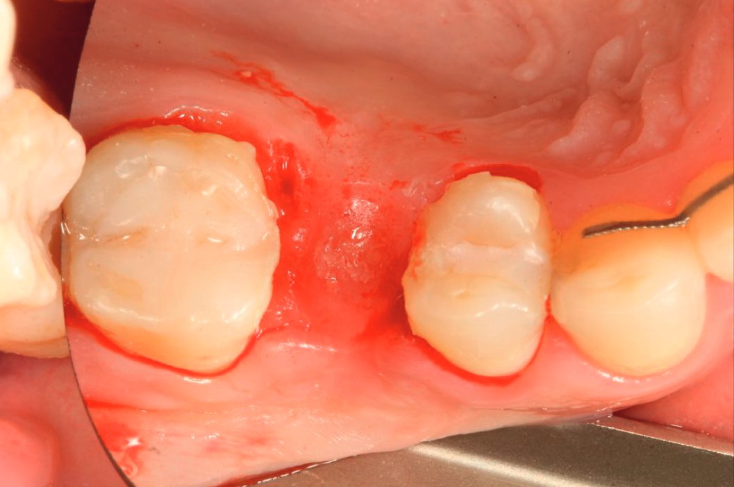
The patient presented three months after ridge augmentation, with ideal healing conditions for implant placement.
Implant placement
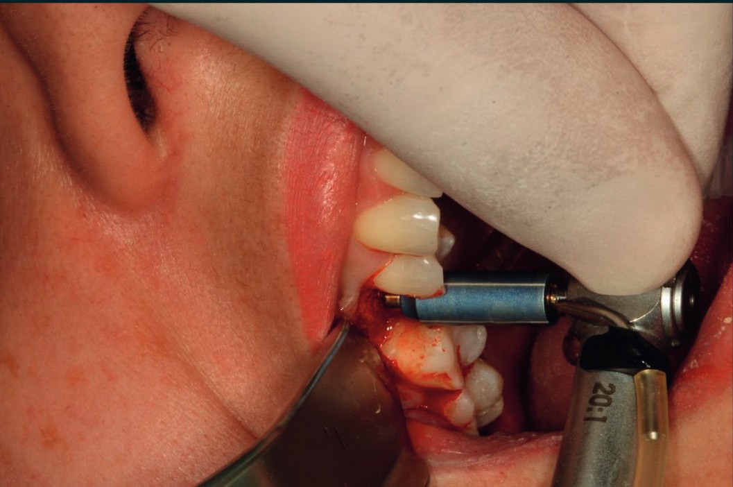
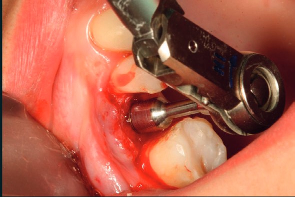
The VPI pilot drill located the exact center of the osteotomy. The PCA implant seated with excellent primary stability from the first moment.
Seating the i-base
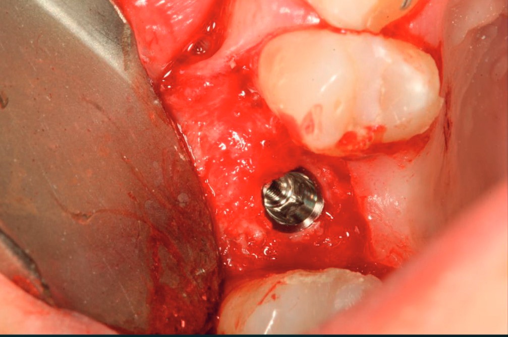
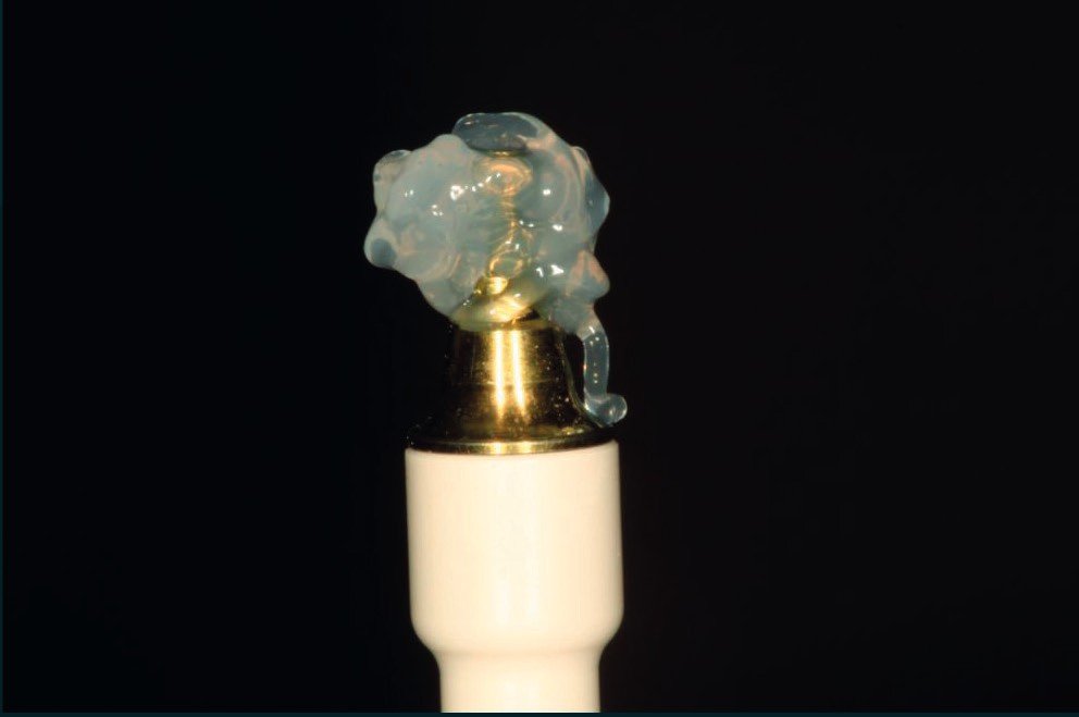
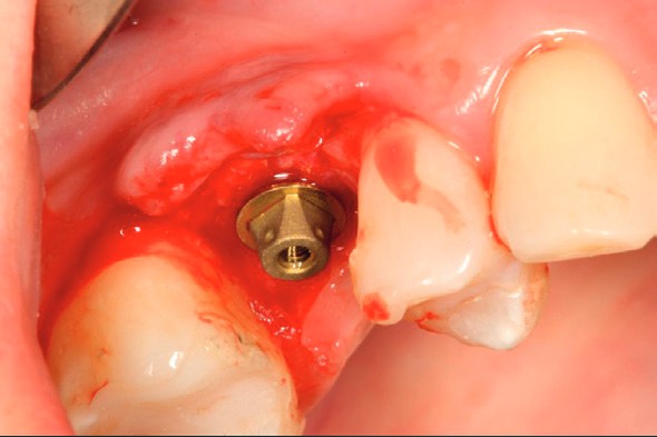
A 1.5 mm cuff-height i-base was selected and coated with antiseptic gel, then seated on the implant and torqued to 35 Ncm. It stays in place from this moment on.
Shaping the soft tissue
A small centered premolar (pS) iCervico healer was placed on the i-base, oriented and adjusted on the distal aspect for optimal space management, and torqued to 25 Ncm. An immediate provisional was then built in resin directly on the healer profile.
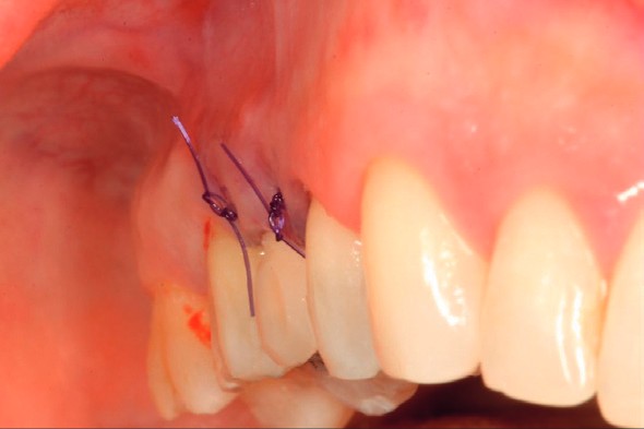
Three weeks later
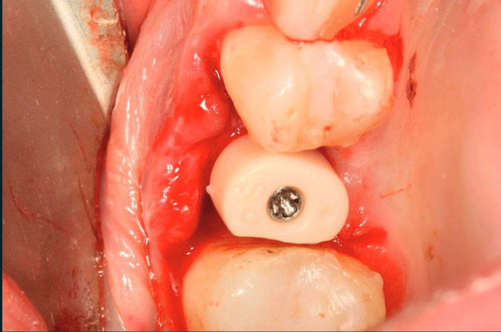
The healer was removed, revealing excellent soft-tissue profile shaping. The AR-sleeve was then placed on the i-base for digital scanning; no transgingival disconnections at any point.
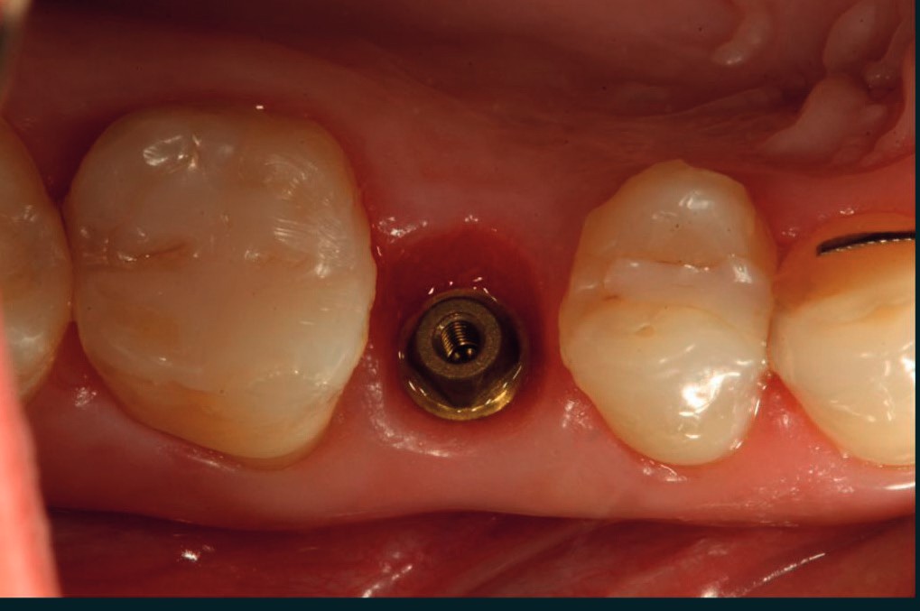
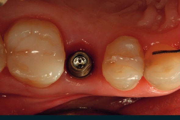
Final restoration
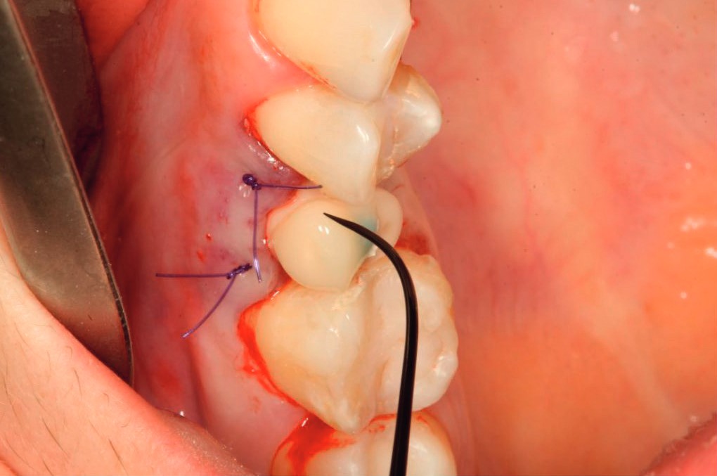
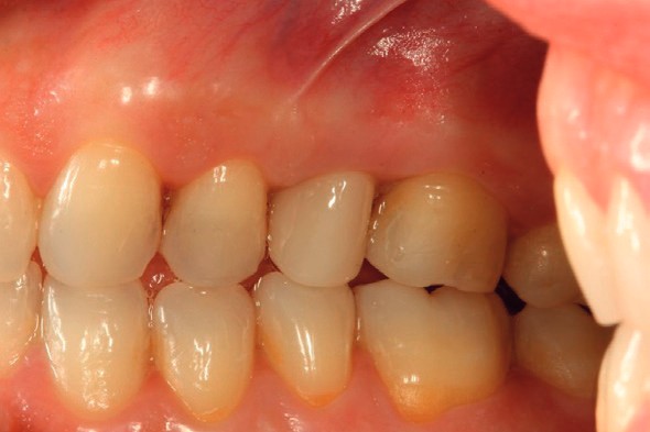
The final prosthesis delivered on the implant showed excellent soft-tissue adaptation, a functionally sound occlusion and excellent aesthetics.
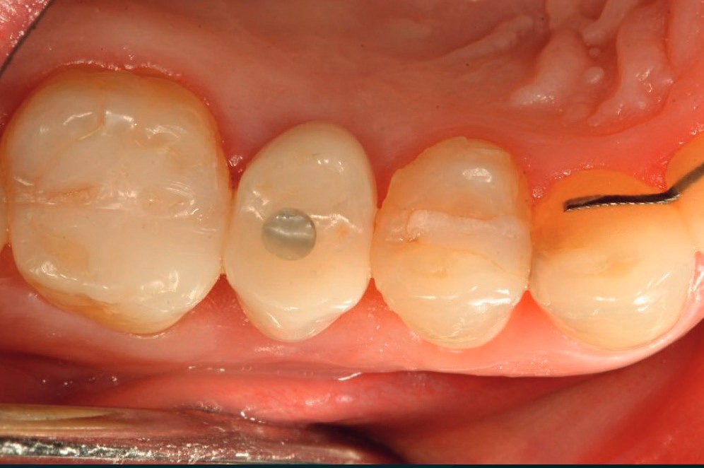
Clinical contributors.
Every component used in this case, from the i-base to the AR-sleeve, is covered in the product brochure.
Dimensions, torque values and ordering references for the full system.
Download the brochure (PDF)